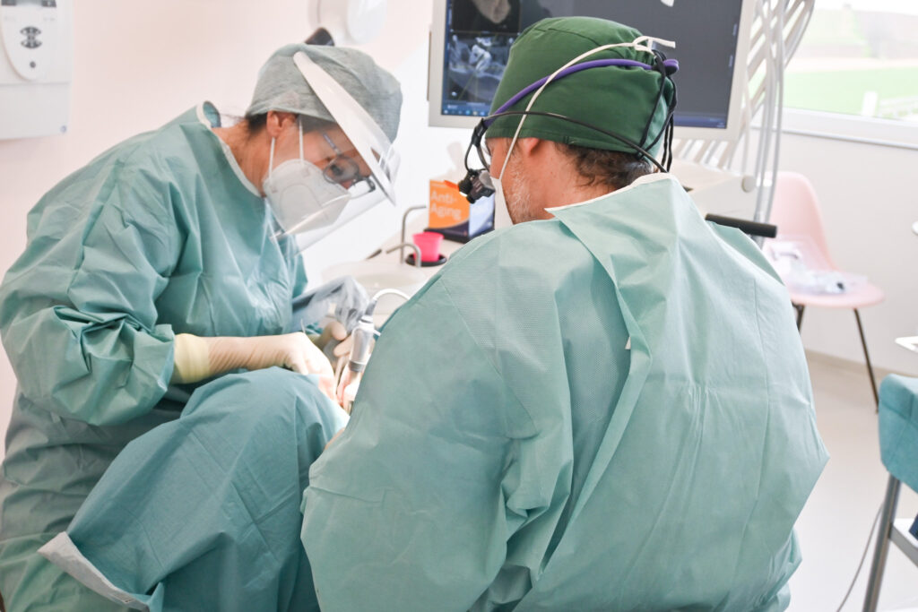
Echocardiography, also known as cardiac ultrasound, is a non-invasive imaging technique used to assess the structure and function of your heart. It is based on the use of ultrasound waves that are generated by a special device and then provide real-time images of your heart. These images allow doctors to examine various aspects of the heart, including the size of the heart chambers, the thickness of the heart walls, the movement of the heart valves and the pumping function of the heart.
Echocardiography is commonly used to diagnose and assess a variety of heart conditions, including heart valve defects, heart muscle thickening (hypertrophy), heart muscle inflammation (myocarditis), congenital heart defects and heart failure. The examination allows doctors to carry out a more precise root cause analysis for symptoms such as shortness of breath, chest pain or irregular heart rhythm.
Types of examination
There are different types of echocardiography, including transthoracic echocardiography (TTE), where the transducer of the ultrasound machine is placed over your chest wall.
Echocardiography is a safe and painless examination that is usually performed on an outpatient basis and requires no special preparation. It provides important information for the diagnosis and management of heart disease and plays an essential role in everyday clinical practice in cardiology.


