Abdominal ultrasound, also known as abdominal sonography, is a diagnostic procedure used to examine your internal organs in the abdomen. This includes organs such as the liver, gallbladder, pancreas, spleen, kidneys and the major blood vessels that run through your abdomen.
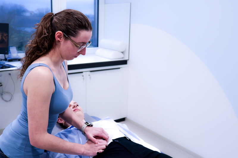
During an abdominal ultrasound scan, you usually lie on your back while the doctor applies a gel to your abdomen to ensure good sound transmission. A hand-held ultrasound device is then moved across your abdominal wall, generating high-frequency sound waves that penetrate your body and are reflected by the various organs.
How does the examination work?
The sound waves generated are converted by a computer in real time into images that are displayed on a screen. The internist can then assess the structure and condition of your examined organs. For example, changes in the size, shape, texture and density of the organs may indicate abnormalities, such as enlargement of the liver, gallstones, tumors or fluid accumulation.
Why is abdominal sonography useful?
Abdominal ultrasonography is a non-invasive and painless procedure that usually requires no special preparation and involves no radiation exposure. It is often used to diagnose a variety of diseases and conditions in the abdomen and can also be used during pregnancy to monitor fetal development.
Due to its safety, accessibility and effectiveness, abdominal ultrasonography is a valuable tool in the diagnosis and monitoring of abdominal diseases and plays an important role in medical care.