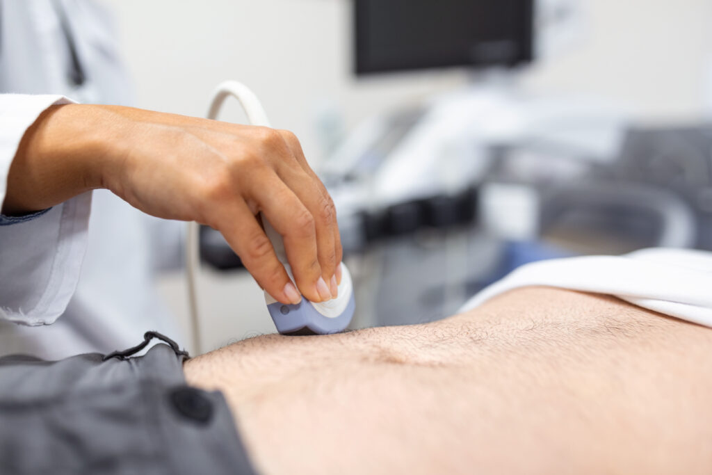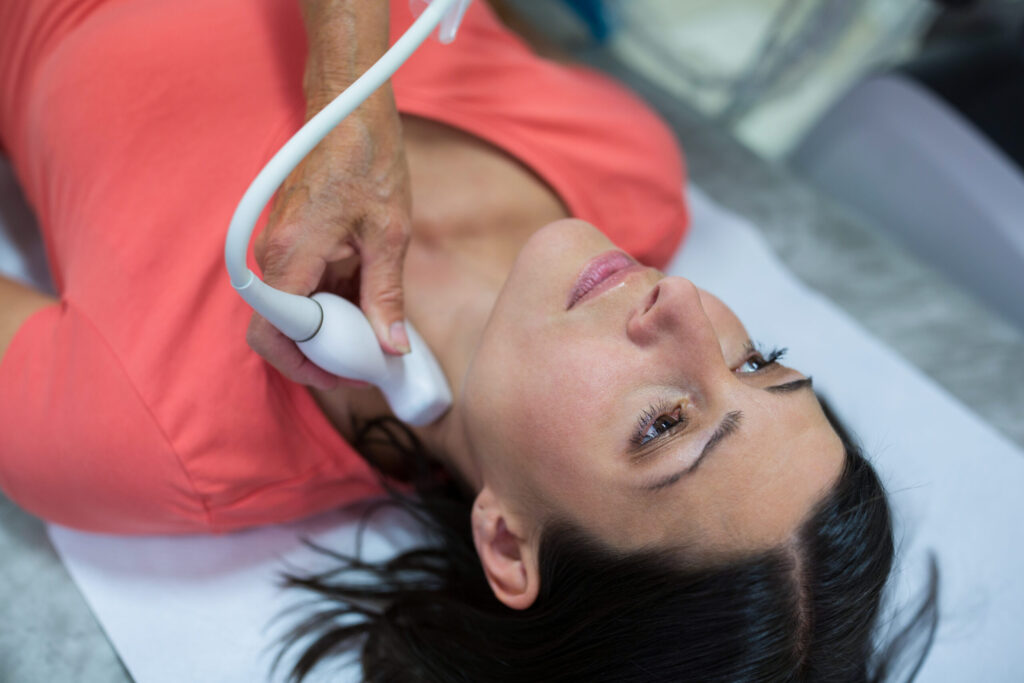
Carotid ultrasonography (ultrasound of the neck vessels) is an imaging procedure used to examine your blood vessels in the neck, particularly the carotid arteries. Carotid ultrasonography, also known as Doppler ultrasonography of the carotid arteries, is an important part of vascular diagnostics. This non-invasive procedure uses ultrasound waves to visualize and evaluate the blood flow in your carotid arteries.
When is this examination used?
Carotid ultrasonography is often used to assess circulatory disorders in the carotid arteries, particularly in cases of suspected arteriosclerosis or narrowing of the arteries (stenosis). These narrowings can lead to an increased risk of strokes, as they can impair the blood flow to the brain.
How does this sonography work?
During this procedure, an ultrasound device is applied to the skin in the area of your neck. By using high-frequency sound waves, the structures of the carotid arteries are visualized and the blood flow is measured using the Doppler effect. This allows doctors to assess the degree of narrowing of the arteries and possible deposits (plaques) in the vessels.
Why is carotid sonography useful?
The results are decisive for further treatment, for example, in the case of vascular diseases. Depending on the findings, drug therapy, interventional treatment such as vascular support (stenting) or surgical removal of the deposits may be necessary to reduce the risk of a stroke.
Overall, carotid sonography is a safe and reliable procedure for assessing the carotid arteries and plays an important role in the early detection and treatment of vascular diseases that can increase the risk of a stroke.